The Development of Neurophysiology
We next turn
to physiology. Though physiology is not
on our list of core disciplines in cognitive science, it has a special
significance in the development of both psychology and neuroscience. Psychology has two parent disciplines;
Philosophy and Physiology. Philosophy
introduces the “big questions” about the nature and operations of the mind. Physiology--particularly the early physiology
of the nervous system--marks the beginnings, not only of neuroscience, but also
of the introduction of experimental methodology to the study of the mind.
The early
development of physiology involves the development of an accurate knowledge of
the gross anatomical structures of the body, and improved understanding of the
functions of anatomical structures, and the development of an experimental
methodology for testing functional hypotheses.
From that point, neurophysiology and eventually neuroscience are fueled
primarily by increasing the repertoire methodologies, both for visualizing ever
finer structures and for experimentation to determine the functions of newly
visualized structures.
As we’ll see,
knowledge of even the gross anatomical structures of the nervous system and their
function eludes theorists throughout most of history. This ignorance has two sources. First, prohibitions and/or stigma around
dissection of human corpses create a lack of empirical investigation and
documentation as to the gross anatomical structures. Second, a lack of valid empirical experimental
methodology provides for very limited understanding of the corresponding
functions and functional organization of gross anatomical structures.
Aristotle (384-322 BCE) performs dissections of animals to gain anatomical knowledge. His anatomical studies lead him to view the brain primarily as a radiator for the heart which generates sensations and emotions in 335 B.C.E.. In Parts of Animals1 Aristotle claims that “...the brain cannot be the cause of any of the sensations, seeing that it is itself as utterly without feeling as any one of the excretions.” (Part 10, ¶2)
 |
| Aristotle (384-322 BCE) |
 |
| Human ventricles of the brain are a system of four structures continuous with the central canal of spinal cord. They are shown in mid-sagittal plane (middle) picture of the right hemisphere (upper left), and (upper right) coronal plane (vertical plane dividing the brain into anterior [front] and posterior [back]) |
While the
Greek physicians Herophilus of Chalcedon (335-280 BCE) and Erasistratus of Chios (304-250 BC) perform relatively
systematic human dissections around 300 BCE.
Even in the enlightened Greek culture, the practice is stigmatized and
in many places illegal. However, in the
third century BC under Ptolemy I and his successor, Ptolemy II Philadelphus,
the city founded by Alexander of Macedonia, Alexandria, develops a relatively
unique social, cultural, political and intellectual climate which allows Herophilus
and Erasistratus the freedom to dissect human bodies. Both before and after the period of
Hellanistic Alexandria physicians looked for the most part to the dissection of
animals to gain knowledge of the human anatomy.
Herophilus suggests that the ventricles are
seat of mentality. It isn’t until the
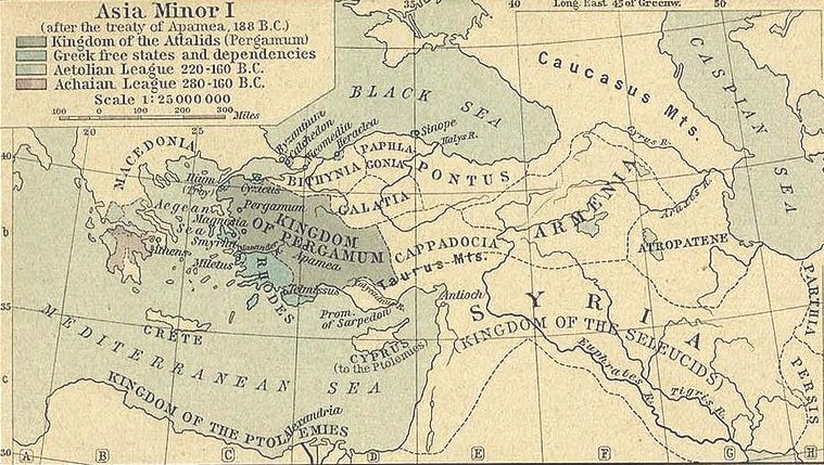
Roman physician Galen of Pergamum delivers a series of lectures in 177 ADE that
any anatomist considers brain itself as the seat of mentality. Galen (Claudius Galenus 129-200ADE), begins his career as Roman
physician in his home town of of
Pergamum, a Greek city, like Miletus, in Asia Minor.
Pergamum is one of the greatest cultural and academic cities of its time,
second only to Alexandria where Galen also studied medicine.
 |
 |
|
Pergamum circa
188BC http://en.wikipedia.org/wiki/File:Asia_Minor_188_BCE.jpg |
(Claudius Galenus 129-200AD) |
 |
|
|
Pineal Gland http://www.vivo.colostate.edu/hbooks/pathphys/endocrine/otherendo/pineal.html |
|
Scholars often describe Galen as the greatest medical
researcher of the Roman period. He makes
significant contributions to medicine, anatomy, physiology, logic and
philosophy that shape thought in these areas for centuries. Roman
law forbids dissection and autopsy of the human body, so Galen, like all
physicians of the times is unable learn from human cadavers. Instead, he dissects the Barbary Macaque and
other primates, transferring what he learns on the assumption that the
anatomies are basically the same.
However, Galen does have an advantage that most anatomists do not. As a physician to gladiators, Galen has many
occasions to view the internal organs of their bodies. In Galen’s major anatomical work, De
Usu Partium Corporis Humani (On
the Usefulness of the Parts of the Body ca. 210 AD) as well as in a famous
earlier lecture (177ADE) he describes the ventricles and pineal
gland. He argues
against the idea--apparently widely espoused at the time--that the ventricles
are filled with “psychic pneuma,” the airy or vaporous substance supposed to
interact with both body and mind. Galen
also argues against the idea that the movements of the pineal gland act to
regulate the flow of these pneuma within the ventricles. No one really listens, and such theories
continue to dominate thinking throughout the middle ages.
The first anatomy textbook published in Europe is Anothomia written by the Italian
physician Mondino de'Luzzi (ca. 1265-1326). Though de’Luzzi completes
the text in 1316 while at the University of Bologna, it is not published until after
his death in 1478 at Padua. The work is
based upon de’Luzzi’s own dissections, though most of the errors in Galen are
repeated. Between the 13th
and 15th century dissection increasingly becomes accepted at universities
throughout Europe, together with an increasing interest in, and knowledge of
anatomy. Interest in the brain remains
centered upon the ventricles. Leonardo
da Vinci (1452-1519) creates a cast of the human ventricles in 1504. In 1536 the italian Niccolò Massa (1485–1569)
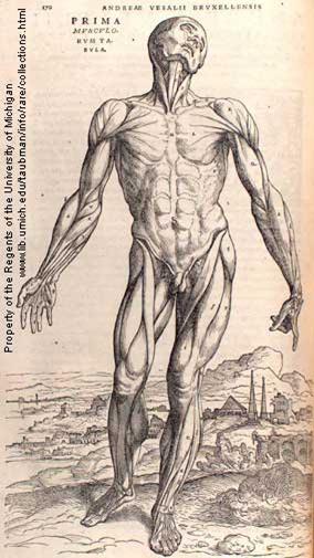
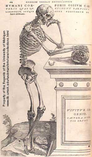
argues that the ventricles are filled with fluid (liquor cerebro-spinalis). Andreas Vesalius (1514-1564), a Belgian physician/anatomist
publishes the next major anatomical work.
Vesalius’ seven volume De
Humani Corporis Fabrica (On the Workings of the Human Body) in
1543. In the volume, Libri septem, of Fabrica, Vesalius argues against all ventral localizations and all
theories that the pineal and other structures regulate the flow of animal
spirits.
 |
 |
| Leonardo da Vinci (1452-1519) | Andreas Vesalius (1514-1564) |
 |
 |
|
Images from Vesalius’ De
Humani Corporis Fabrica http://vesalius.northwestern.edu/flash.html |
|
Vesalius’ work represents a dramatic improvement upon previous
anatomical knowledge, and corrects many of the mistakes in Galen. Throughout the remainder of the 15th
century anatomists continue to improve their knowledge of anatomical
detail. In 1550 Bartolomeo Eustachio (1514-1574),
an Italian contemporary of Vesalius, traces the optic nerves to their origins
in the brain. Gabriele Falloppio’s Observationes Anatomicae (1561) includes
a description of some of the cranial nerves.
Giulio Cesare Aranzi (1529-1589) coins the term hippocampus in his De Humano Foetu Opusculum (1564). Girolamo Mercuriali (1530-1606) writes De Nervis Opticis in 1573 which further
documents optic nerve anatomy.
Aranzi also stands out in the development of experimental methodology. His Observationes Anatomicae (1587) introduces the results of his experimental work on anatomical function, including his demonstration that the retina has a reversed image. Other contributions to a functional understanding of the nervous system include a work on neurological disease by Jason Pratensis (1486-1558) called De Cerebri Morbis (1549). Vesalius, who is working as a private physician at the time, publishes Hydrocephalus ex vacuo (1550), which provides an accurate account of hydrocephalus (a build-up of fluid inside the skull, leading to brain swelling).
 |
 |
| Girolamo Mercuriali (1530-1606) | Johannes Kepler (1571-1630) |
 |
 |
| Christoph Scheiner (1573-1650) | Jason Pratensis (1486-1558) |
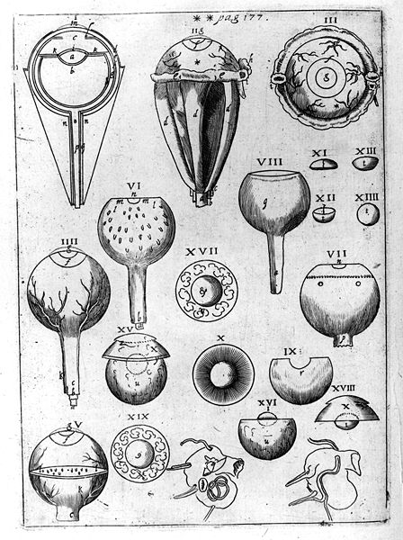 |
A plate (left) from Kelper’s Ad Vitellionem paralipomena, quibus Astronomiae pars optica traditur depicting the structure of the eye. From http://en.wikipedia.org/wiki/File:Kepler_Optica.jpg |
Christoph Scheiner (1573-1650) another German astronomer
publishes Oculus, hoc est: Fundamentum
opticum (1619). Oculus makes several notable contributions. Scheiner provides the first demonstration
that accommodation is
an active process. He likewise
identifies the retina as the organ of vision.
He is the first to use fixatives to preserve the eye for anatomical
study, which allows him to create the first accurate diagrams of the human eye.
He also discovers the near
pupil reflex.
Accommodation is
a reflexive action by the eye allowing a person to switch their gaze between
objects at different distances. In
order, for example, to switch from viewing a nearby object to viewing a distant
object the eye must adjust the shape of its lens, alter its pupil size (near
pupil reflex), and coordinate its movements with the other eye so as to
maintain the position of the object in the center of the retinas of each
eye. The coordinated movement of the
eyes is called vergence, and it makes binocular depth perception possible.
Scheiner publishes Rosae Ursinae Sive Solis. in 1626-1630. Though these volumes primary concern Scheiner’s observations of the sun, they also include the first direct observation of the retinal image.
 |
  |
| René Descartes (1596-1650) |
Descartes’ diagrams depicting the pineal gland facilitating visual-motor function (left) and the communication of pain (right) in Meditations Métaphysiques |
 |
|
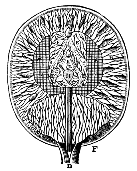
| A diagrammatic section of human brain from René Descartes’ Treatise on Man | Descartes’ diagram depicting visual processing from La Dioptrique |
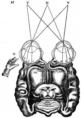
In 1637 René Descartes publishes La Dioptrique as one of three appendices to his Discourse on Method, each offered as an
illustration of Descartes’ method. Dioptrique is a treatise on optics. Though not particularly original in its
results from optics, it articulates the corpuscular theory of light and suggests
for the first time that the retina projects directly onto brain (in his view,
onto the walls of the ventricles).
Though a dualist, Descartes makes some of the
first steps towards psychology and neuroscience. Descartes maintains a very strong long-term interest
the workings of the physical body, and spends a great deal of time dissecting
cadavers. His theory of mind-body
interaction is based upon my knowledge of gross neuroanatomy. Specifically, (1)
Descartes posits the pineal gland as the “seat” of mind-body interaction. He hypothesizes, contra Galen, that the pineal
gland plays a role in sensation, imagination, memory and the causation of
bodily movements as early as my first work, Treatise
of Man (written 1637, published 1662). Thus, the pineal gland serves as the
principle organ for sensus communis--the communication between the body and the
soul. Both the soul and the body’s animal
spirits can affect the pineal gland by literally moving it, thereby allowing
each to act on the other. Additionally, (2) Descartes adopts Galen’s hypothesis that the
nerves are hollow tubes that contain2 “…a certain very
subtle wind, or rather a very lively and pure flame, which is called ‘animal
spirits’.” (p.19)
Descartes’ view proves important, in part because it becomes very influential. It is also based on physiology--though he am mistaken on numerous points regarding the pineal gland, etc.. Finally, Descartes views the body as a machine capable of autonomous action--thereby indirectly furthering physical explanations of the mind. Indeed, he notes that,2
|
…it is not necessary to conceive of this machine as having any vegetative or sensitive soul or other principle of movement and life, apart from its blood and its spirits, which are agitated by the heat of the fire burning continuously in its heart—a fire which has the same nature as all the fires that occur in inanimate bodies. (p.113) |
We’ll end this section with a final contribution from
optics. Robert Hooke (1635-1703)
is a somewhat obscure English “natural philosopher” and a polymath. Nevertheless, he proves remarkably
influential in the sciences, through both his experimental and his theoretical
contributions. Hooke’s Law, which
describes the tendency of elastic materials to deform under stress, probably
stands out as Hooke’s best-known scientific contribution. However, from the perspective of cognitive
science, Hooke’s legacy derives from his position as the “Father of
Microscopy.”
 |
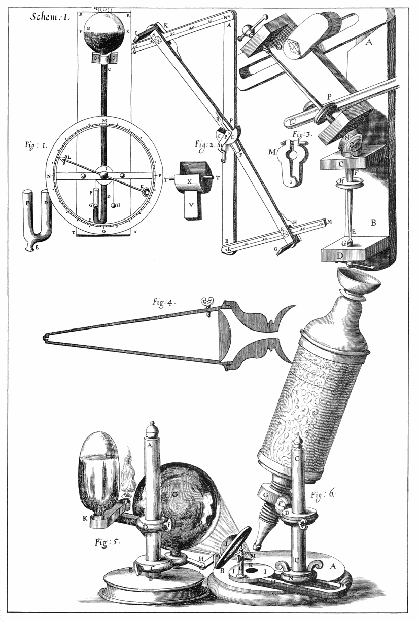 |
| Robert Hooke (1635-1703) | Hooke’s diagram of his microscope from Micrographia |
 |
 |
| Hooke’s microscope | Hooke’s drawing of cork cells from Micrographia |
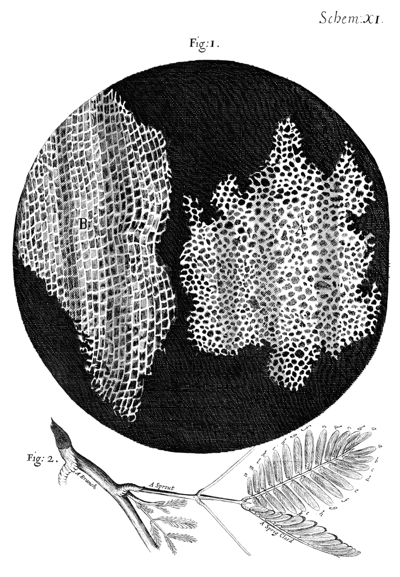
Hooke publishes Micrographia
in 1665. The book contains a record of Hooke’s
observations using a microscope (of his own devising). Hooke’s observations of cork, specifically,
the presence of cells (see drawing), prove the most significant from the
standpoint of cognitive science. One
cannot underestimate the existence of visualizable cells, in the development of
biology and neurobiology. Hooke himself,
however, did not observe cells in animals, and did not believe that animals
were composed of cells.3
|
First, in that it had a
very little solid substance, in comparison of the empty cavity that was
contain'd between, as does more appear by the Figure A and B of the XI. Scheme,
for the Interstitia, or walls (as I may so call them) or partitions of
those pores were neer as thin in proportion to their pores, as those thin films
of Wax in a Honey-comb (which enclose and constitute the sexangular celts)
are to theirs. Next, in that these pores, or cells, were not very deep, but consisted of a great many little Boxes, separated out of one continued long pore, by certain Diaphragms, as is visible by the Figure B, which represents a sight of those pores split the long-ways. (p.113) |
Ending the discussion of physiology here is somewhat
arbitrary. Few historians suggest a
specific time for the beginning of neuroscience. However, we will return to the scientific
study of the brain when we discuss neuroscience. The discussion in this section focuses upon
the development of gross anatomical and neuroanatomical knowledge, the introduction
of experimental techniques and devices in physiology, and the early attempts at
identification of the function of gross neuroanatomical structures. These three developments eventually allow
theorists to begin to systematically study the nervous system at the many
levels of functioning that characterize neuroscience.
Sources
| 1. | Aristotle. On the Parts of Animals (MIT Internet Classics Archive, 350 BCE). |
| 2. | Descartes, R. Traite de l'homme (Treatise on Man) (ed. Hall, T.S.) (Prometheus Books, Amherst. NY, 2003). |
| 3. | Hooke, R. Micrographia: or, Some physiological descriptions of minute bodies made by magnifying glasses (J. Martyn and J. Allestry, London, 1665). |